Principle
Optical microscopy provides color images at high magnification of the surface states, the corrosion processes, paint layers …
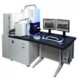
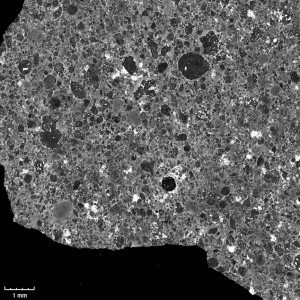
The SEM provides black and white images of the studied matter at very high magnification, using an electron beam and electromagnetic fields.
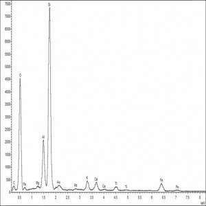
Furthermore, the electron energy used is sufficient to allow interactions with the atoms of the material giving rise to spectra providing information about the elemental composition of the analyzed material (XRF).
Measurements
They are carried out on micro-samples, mostly embedded in a polymer resin to ensure a polished optical surface. The results, both in terms of imaging and elemental analysis, constitute a useful set of data to characterize the material and to validate the compatibility of its composition and highlighted weathering processes with the presumed antiquity of the object. This approach works really well on inorganic materials such as stones, metals, glazes materials …
On organic materials such as wood, ivory, fabrics … we observe their textures and variations.
 |
 |
 |
Download the document about microanalysis on bronze![]()
Download the document about microanalysis on stones![]()
Any question ? Any thought you would like to share ?
Contact us